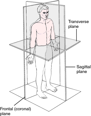
I have some nii.gz files which, when I open using imajeJ , or any python library such as SimpleITK or Monai I get a stack of MRI images, as if taken from top to bottom. That being said, when I open the same file with slicer, I also get a stack of images, taken from the side and from the front, the G and Y channels. My question is, do the images from the different angles exist inside the file, or is 3D slicer inferring how these channels would look like based on the R channel. I am asking this because the other methods don’t seem to be able to find the other views. Thank you.
3D slicer

ImageJ

Monai. SimpleITK in google colab
