Patent ductus arteriosus
Background
- Ductus arteriosus is a blood vessel connecting main pulmonary artery to proximal descending aorta
- Shunts blood from right ventricle to bypass fetus's non-functioning lungs in utero
- In PDA, ductus fails to close after birth
- Second most common congenital heart disease
- Approximately 1 in 1600 to 5000 live births in the US.[1]
Pathophysiology

- Ductus arteriosus shunts blood from the pulmonary artery to the aorta bypassing the lungs while in utero
- Normally closes soon after birth via complex process
- Regulated by oxygen tension and decreases in prostaglandin E2
- Becomes ligamentum arteriosum
- In PDA, ductus fails to close
- Results in shunting of blood between aorta and pulmonary artery
- Some oxygenated blood from high-pressure aorta shunts to lower pressure pulmonary artery
- A large shunt can cause increased fluid return to lungs and increased lung pressure, leading to pulmonary hypertension
- Some congenital heart defects, such as transposition of the great arteries, are incompatible with life 'without a PDA; may be necessary to allow oxygenated and deoxygenated blood to mix
- In these cases prostaglandins are used to keep the ductus arteriosus open.
Clinical Features
- Continuous machine like heart murmur
- Differential cyanosis (cyanosis of lower extremities and not the upper extremities)
- Dyspnea
- Tachycardia
- Widened pulse pressure
- Congestive heart failure
- Failure to thrive
Differential Diagnosis
- Acute pericarditis
- Aortopulmonary Septal Defect
- Coarctation of the Aorta
- Coronary artery fistula
- Pediatric acute respiratory distress syndrome
- Sinus of valsalva aneurysm
- Pediatric tachycardia
- Pulmonic valvular stenosis
- Sickle cell anemia
- Tetralogy of Fallot with absent pulmonary valve
Congenital Heart Disease Types
- Cyanotic
- Acyanotic
- AV canal defect
- Atrial septal defect (ASD)
- Ventricular septal defect (VSD)
- Cor triatriatum
- Patent ductus arteriosus (PDA)
- Pulmonary/aortic stenosis
- Coarctation of the aorta
- Differentiation by pulmonary vascularity on CXR[2]
- Increased pulmonary vascularity
- Decreased pulmonary vascularity
- Tetralogy of fallot
- Rare heart diseases with pulmonic stenosis
Evaluation
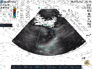
Echocardiography demonstrating PDA[3]
- Careful physical examination demonstrating characteristic machine like murmur
- Echocardiography
- MRA and Cardiac CT can also be used as diagnostic tools
- Laboratory tests usually not helpful
Management
- Spontaneous closure common
- If significant respiratory distress or impaired oxygen delivery, therapy usually required
Medical Therapy
- IV indomethacin: usually effective when administered in the first 10-14 days of life.
- <48 hour old: start 0.2mg/kg IV x 1, then 0.1mg/kg q12-24h x 2
- 2-7 days old: Start 0.2mg/kg IV x 1, then 0.2mg/kg q12-24h x 2
- > 7 days old: Start 0.2mg/kg IV x 1, then 0.25mg/kg q12-24h x 2
Surgical Therapy
- Offered if medical therapy fails
- Cardiac catheterization and catheter closure
- Surgical ligation
Disposition
- Stable, asymptomatic patients can be discharged home with cardiology follow-up
- Symptomatic patients and patients requiring treatment should be admitted with cardiology consultation
See Also
- Congenital Heart Disease
External Links
References
- Mitchell, S. C., Korones, S. B., Berendes, H. W. Congenital heart disease in 56,109 births: incidence and natural history. Circulation 43: 323-332, 1971.
- Knipe K et al. Cyanotic congenital heart diseases. Radiopaedia. http://radiopaedia.org/articles/cyanotic-congenital-heart-disease
- http://www.thepocusatlas.com/pediatrics/
This article is issued from Wikem. The text is licensed under Creative Commons - Attribution - Sharealike. Additional terms may apply for the media files.